The Cardiac Skeleton Is Best Described as __________________ .
Single nucleus and spindle-shaped cells. Also skeleton of the heart reinforced connective tissue located within the atrioventricular septum.

Lec 10 Flashcards Quizlet
Structure of the Heart.

. The skeleton in 30 otter hearts was studied by x-ray analysis and light microscopy. Understood as such the cardiac skeleton efficiently centers and robustly funnels electrical energy from the atria to the ventricles. A bag of bones.
This muscle tissue which contracts and releases involuntarily is. Provides electrical insulation between the atria and ventricles. Which of the following statements best describes the changes that occur in the skeletal system as a person ages.
Dense bands of tough elastic tissue that encircle the heart valves The coronary arteries emerge at the base of the __________. The internal framework of the body. The period of timethat begins with contraction of the atria and ends with ventricular relaxation is known as the cardiac cycle Figure 1931.
The size and location of each oval show where the structure belongs in the levels of organization. The period of contraction that the heart undergoes while it pumps blood into circulation is called systole. The atrioventricular conducting system is the only electrical connection between the atria and the ventricles in a normal heart.
A collection of two or more types of tissue that work together to perform a specific function is called a n What are the five levels of organization in your body. Asked Nov 28 2021 in Biology Microbiology by Fantastic_One. The upper limbs are held out to each side and the palms of the hands face forward as illustrated in Figure 141.
It plays an important role in making your heart beat. The cardiac skeleton also known as the fibrous skeleton of the heart is a high density singlehomogeneous structure of connective tissue that forms and anchors the valves and influences the forces exerted by and through them. Layers of the Heart Wall.
Multinucleate and long cylindrical cells. E More questions like this The nurse is caring for a patient with skeletal pins that have been placed for traction. The cardiac skeleton separates and partitions the atria the smaller upper two chambers from the ventricles the larger lower two.
It reinforces the myocardial walls internally and anchors the cardiac muscle. Which of these characteristics best describes cardiac muscle tissue. The heart is an organ with walls composed of cardiomyocytes supported by fibrocollagenous tissue.
Attached to the skeleton. Which of the following best describes the human skeleton. Using this standard position reduces confusion.
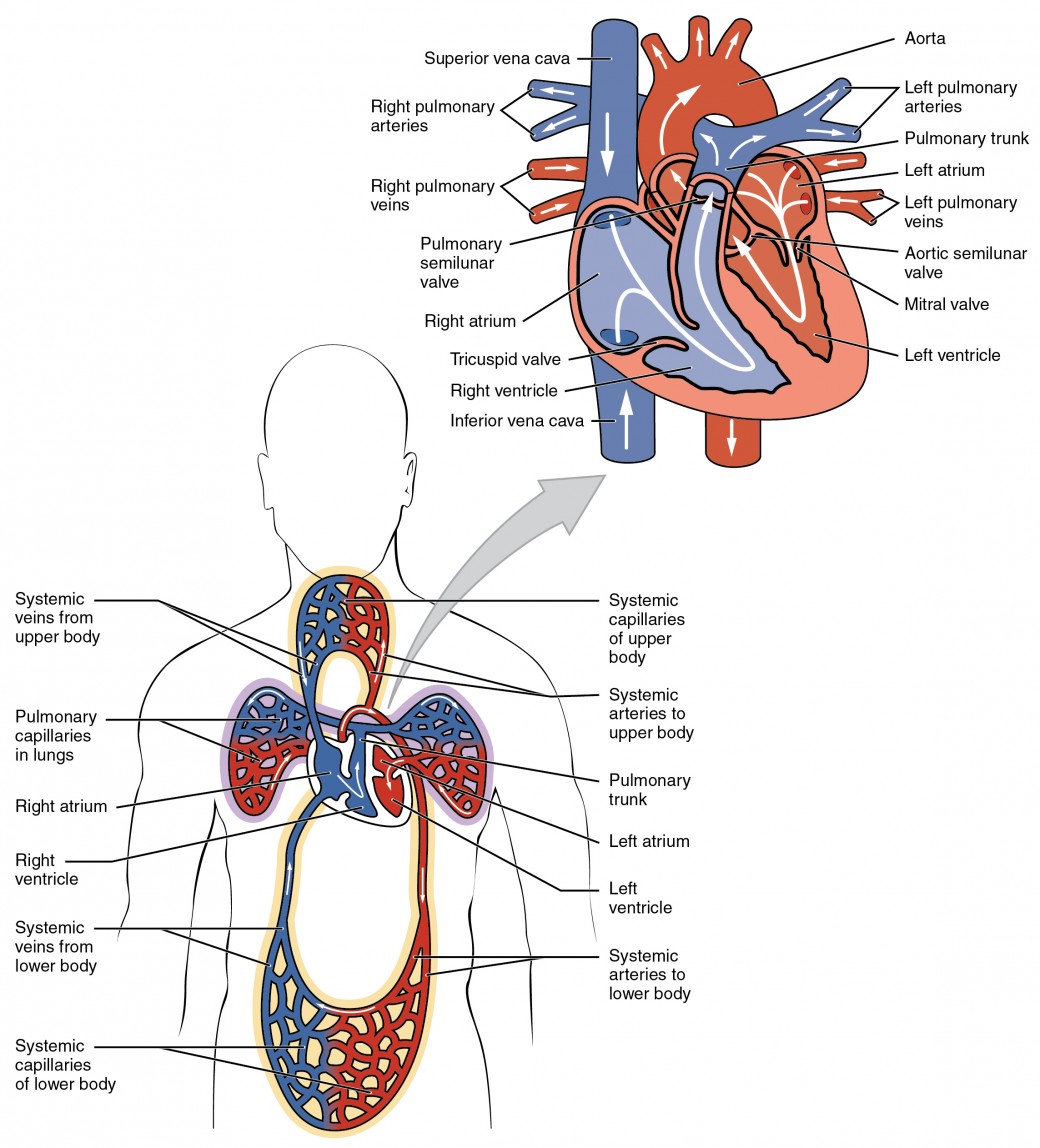
The heart is enclosed in a pericardial sac that is lined with the parietal layers of a serous membraneThe visceral layer of the serous membrane forms the epicardium. Well go over the unique features of cardiac muscle tissue. Dense bands of tough elastic tissue that encircle the heart valves A large vein that opens into the right atrium and bring venous blood from the heart tissue is the ________.
Skeletal Muscle It is short in length It has a semi-spindle shape Muscle cells are connected side-by-side and are not tightly bound Contractions of muscle cells are interdependent Gap junctions are present in cardiac muscle cells Muscle cell has only 1-2 nuclei Has a dense endomysium. Asked Sep 27 2016 in Anatomy Physiology by Arnold. The cardiac frame is composed of a dense connective tissue network that forms a fibrous skeleton.
How are cells related. The cardiac skeleton of the otter Lutralutra has not previously been described. The cardiac skeleton is best described as _____.
Some of the muscle attached to the skeleton is voluntary and may be used for movement. It reinforces the myocardium it is made up of connective tissue fibers it anchors the cardiac muscle fibers it limits spread of action potentials between certain parts of the heart it forcefully contracts to eject. Includes four rings that surround the openings between the atria and ventricles and the openings to the pulmonary trunk and aorta.
Relate heart sounds detected by auscultation to action of hearts valves. Each oval in this Venn diagram represents a structure in the human body. Which of the following best describes a tissue.
Which of the following is a function of the heart skeleton. Voluntary movement and cells striations. The thickness of the collagen and elastin fibers of the anulus fibrosus varies from area to area and consists of four rings two trigones and one ligament.
A strong structure that makes minerals and oxygen. The exact position of this. Which best describes what this means in terms of movement.
Cardiac muscle tissue is one of the three types of muscle tissue in your body. For example a scar in the anterior front carpal wrist. Cardiac muscle tissue or myocardium is a specialized type of muscle tissue that forms the heart.
The fibrous skeleton of the heart is a connective tissue frame which functions to electrically isolate the atria from the ventricles. The control center of the body. Some of the muscle attached to the skeleton is involuntary and may be used for movement.
Cardiac skeleton anatomy You have 3 more open access pages. The physiologic cardiac skeleton manages to form a firewall governing autonomicelectrical influence until bordering the His Bundle which further governs autonomic flow to the bundle branches of the ventricles. The cardiac skeleton is best described as _____.
QUESTION 25 All of the following are true about the fibrous skeleton of the heart EXCEPT. In this figure oval A represents the digestive system and oval C represents the smooth muscle layer of the stomach. Anatomy and Physiology questions and answers.
It does not matter how the body being described is oriented the terms are used as if it is in anatomical position. Supplies support and reinforcement for the valves. The fibrous tissue is condensed in certain areas to form a framework often called the fibrous skeleton which anchors the valvar leaflets to the base of the ventricular mass.
The aortic valve occupies the central position with the other valve rings attached. Any muscle attached to the skeleton is voluntary and may be used for movement. The cardiac skeleton consists of four rings of dense connective tissue that surround the AV canals mitral and tricuspid and extend to the origins of the aorta and the pulmonary trunk providing structure and support for the heart as well as electrical isolation between the atria and the ventricles.
Involuntary movement and cell striations. The human heart is a four-chambered muscular organ shaped and sized roughly like a mans closed fist with two-thirds of the mass to the left of midline.

Lec 10 Flashcards Quizlet

Anatomy And Physiology Of The Heart Springer Publishing

Anatomy And Physiology Of The Heart Springer Publishing

Lec 10 Flashcards Quizlet

Anatomy And Physiology Of The Heart Springer Publishing
Heart Anatomy Anatomy And Physiology

Cardiac Muscle And Electrical Activity Anatomy And Physiology Ii

Heart Anatomy Anatomy And Physiology

Heart Anatomy Anatomy And Physiology

Anatomy And Physiology Of The Heart Springer Publishing

Lec 10 Flashcards Quizlet

Lec 10 Flashcards Quizlet

Lec 10 Flashcards Quizlet
Section 46 3 Muscular System

Heart Anatomy Anatomy And Physiology

Heart Anatomy Anatomy And Physiology

Heart Anatomy Anatomy And Physiology

Chapter 18 The Cardiovascular System Flashcards Quizlet
Anatomy Heart Ch 11 Mastering A And P Chapter 19 Homework Blood Vessels Blood Pressure And Vessels Mastering A P Practice Questions Chapter 19 Mastering Anatomy Chapter 19 Blood Vessels A P Cardiovascular Chpt


Comments
Post a Comment